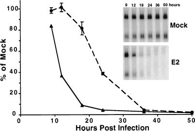
Figure 1.
Time course of HPV E6/E7 repression and growth inhibition. HeLa cells were infected or mock-infected, and, at the indicated time after infection, RNA was analyzed for HPV E6/E7 expression by Northern blotting (solid line), and cellular DNA synthesis was determined by incorporation of tritiated thymidine (dashed line). The error bars indicate two standard deviations of the mean. (Inset) Repression of HPV E6/E7 expression. Northern blot described above. RNA was isolated at the indicated hours after E2 infection (Lower) or mock-infection (Upper), electrophoresed, transferred, and probed with a radiolabeled HPV18 E6/E7 DNA fragment. The signal obtained was quantitated, normalized for the signal obtained with a ubiquitin probe, and expressed as the percentage of the normalized signal obtained with RNA from mock-infected cells.

