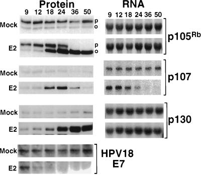
Figure 4.
(Left) Western analysis of retinoblastoma family members. HeLa cell protein was prepared at the indicated hours after E2 infection or mock-infection. After electrophoresis and transfer, specific antibodies were used to detect HPV18 E7, p105Rb, p107, and p130. The hyperphosphorylated (p) and hypophosphorylated (o) form of p105Rb are indicated. (Right) Northern analysis of retinoblastoma family members. HeLa cell RNA was prepared at the indicated hours after mock-infection or E2 infection. After electrophoresis and transfer, p105Rb, p107, and p130 mRNA were detected by hybridization to the appropriate radiolabeled cDNA probe.

