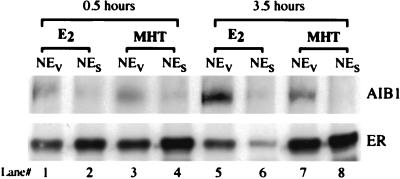
Figure 2.
Coimmunoprecipitation of AIB1 with hER in MCF-7 cells after E2 and MHT treatments. Cells were switched to media with 5% dextran-coated charcoal-stripped FBS for 18 h to withdraw any estrogen. After 0.5 or 3.5 h of ligand treatment, nuclei were extracted with NaCl (NEs) or DV (NEv). Shown is the Western blot analysis for AIB1 (Top) and hER (Bottom) of nuclear extract immunoprecipitations with anti-hER antibody performed as in Fig. 1.

