Abstract
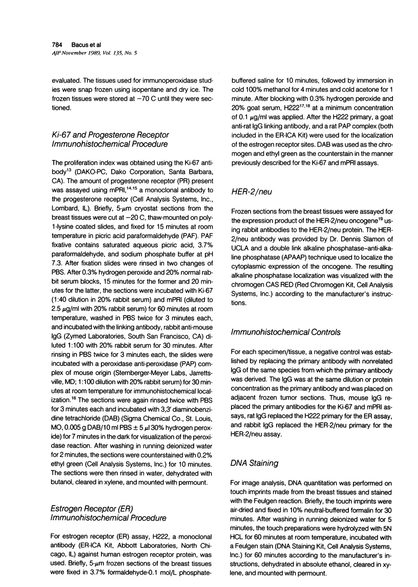
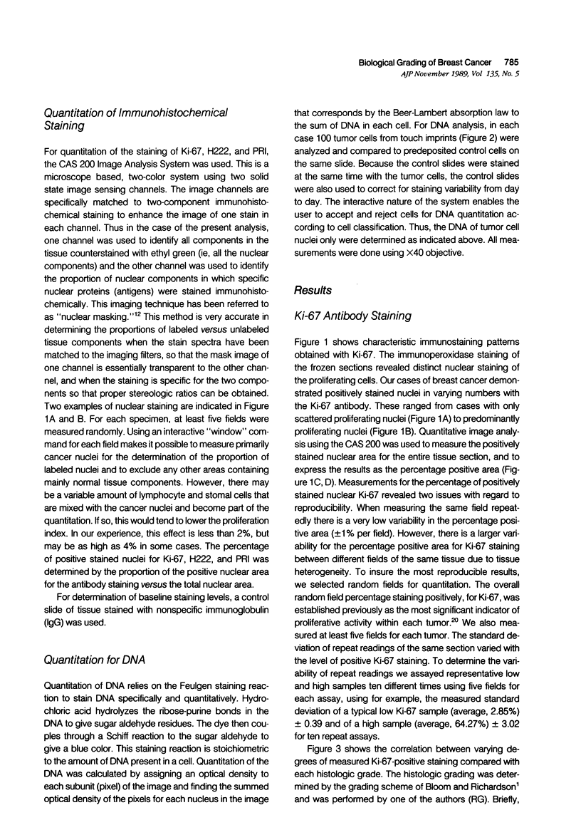
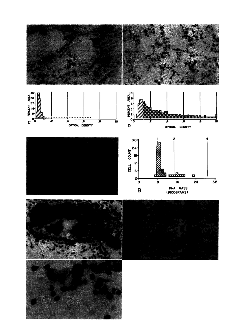
Quantitation of immunohistochemical staining by image analysis was performed on 50 breast cancers stained with the monoclonal antibody Ki-67 to determine the growth fraction and its correlation with tumor grade. A high degree of correlation was shown. For each case the DNA ploidy was determined by quantitation of the DNA Feulgen stain by computerized microdensitometry. DNA content of breast tumor cells correlated to the histopathologic grade at which poorly differentiated tumors are more likely to be aneuploid. Quantitation of immunohistochemistry for estrogen and progesterone receptors had a high degree of correlation with the steroid binding assay, such as dextran-coated charcoal assay (DCCA), and were weakly correlated to histologic grade. In summary, our results indicated that quantitation of Ki-67-positive nuclear area and of DNA content by image analysis provides an objective method for assessing tumor cell growth fraction and DNA ploidy. Quantitation of steroid receptors by immunohistochemistry is a better and easier technique than those currently used to determine the best therapy for postmenopausal women. These methods can be performed on small frozen sections or needle aspirates in quantities that are insufficient for current steroid binding assays. Thus, this method is prognosticly useful even for patients with small breast lesions.
Full text
PDF
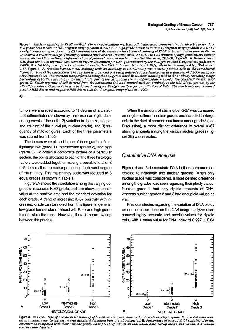
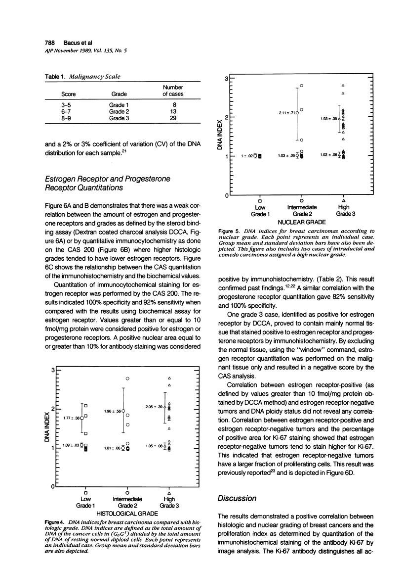
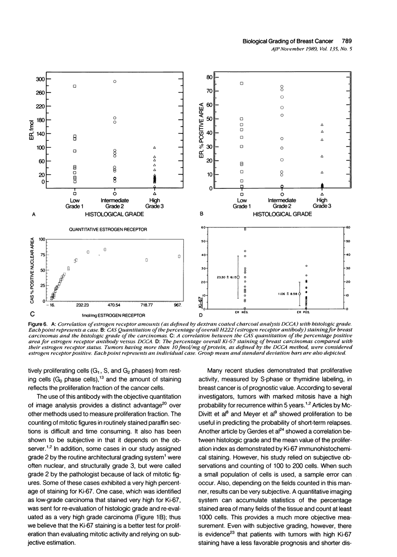
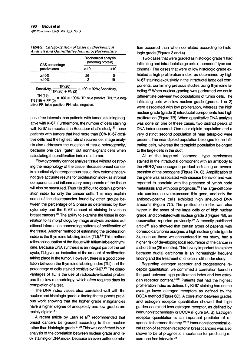
Images in this article
Selected References
These references are in PubMed. This may not be the complete list of references from this article.
- Auer G. U., Fallenius A. G., Erhardt K. Y., Sundelin B. S. Progression of mammary adenocarcinomas as reflected by nuclear DNA content. Cytometry. 1984 Jul;5(4):420–425. doi: 10.1002/cyto.990050420. [DOI] [PubMed] [Google Scholar]
- Auer G., Eriksson E., Azavedo E., Caspersson T., Wallgren A. Prognostic significance of nuclear DNA content in mammary adenocarcinomas in humans. Cancer Res. 1984 Jan;44(1):394–396. [PubMed] [Google Scholar]
- BLOOM H. J., RICHARDSON W. W. Histological grading and prognosis in breast cancer; a study of 1409 cases of which 359 have been followed for 15 years. Br J Cancer. 1957 Sep;11(3):359–377. doi: 10.1038/bjc.1957.43. [DOI] [PMC free article] [PubMed] [Google Scholar]
- Bacus S., Flowers J. L., Press M. F., Bacus J. W., McCarty K. S., Jr The evaluation of estrogen receptor in primary breast carcinoma by computer-assisted image analysis. Am J Clin Pathol. 1988 Sep;90(3):233–239. doi: 10.1093/ajcp/90.3.233. [DOI] [PubMed] [Google Scholar]
- Bauer T. W., O'Ceallaigh D., Eggleston J. C., Moore G. W., Baker R. R. Prognostic factors in patients with stage I, estrogen receptor-negative carcinoma of the breast. A clinicopathologic study. Cancer. 1983 Oct 15;52(8):1423–1431. doi: 10.1002/1097-0142(19831015)52:8<1423::aid-cncr2820520815>3.0.co;2-o. [DOI] [PubMed] [Google Scholar]
- Cornelisse C. J., Van Driel-Kulker A. M. DNA image cytometry on machine-selected breast cancer cells and a comparison between flow cytometry and scanning cytophotometry. Cytometry. 1985 Sep;6(5):471–477. doi: 10.1002/cyto.990060512. [DOI] [PubMed] [Google Scholar]
- Cornelisse C. J., van de Velde C. J., Caspers R. J., Moolenaar A. J., Hermans J. DNA ploidy and survival in breast cancer patients. Cytometry. 1987 Mar;8(2):225–234. doi: 10.1002/cyto.990080217. [DOI] [PubMed] [Google Scholar]
- Fallenius A. G., Auer G. U., Carstensen J. M. Prognostic significance of DNA measurements in 409 consecutive breast cancer patients. Cancer. 1988 Jul 15;62(2):331–341. doi: 10.1002/1097-0142(19880715)62:2<331::aid-cncr2820620218>3.0.co;2-8. [DOI] [PubMed] [Google Scholar]
- Gerdes J., Lelle R. J., Pickartz H., Heidenreich W., Schwarting R., Kurtsiefer L., Stauch G., Stein H. Growth fractions in breast cancers determined in situ with monoclonal antibody Ki-67. J Clin Pathol. 1986 Sep;39(9):977–980. doi: 10.1136/jcp.39.9.977. [DOI] [PMC free article] [PubMed] [Google Scholar]
- Gerdes J., Schwab U., Lemke H., Stein H. Production of a mouse monoclonal antibody reactive with a human nuclear antigen associated with cell proliferation. Int J Cancer. 1983 Jan 15;31(1):13–20. doi: 10.1002/ijc.2910310104. [DOI] [PubMed] [Google Scholar]
- Kamel O. W., Franklin W. A., Ringus J. C., Meyer J. S. Thymidine labeling index and Ki-67 growth fraction in lesions of the breast. Am J Pathol. 1989 Jan;134(1):107–113. [PMC free article] [PubMed] [Google Scholar]
- King W. J., DeSombre E. R., Jensen E. V., Greene G. L. Comparison of immunocytochemical and steroid-binding assays for estrogen receptor in human breast tumors. Cancer Res. 1985 Jan;45(1):293–304. [PubMed] [Google Scholar]
- Lagios M. D., Margolin F. R., Westdahl P. R., Rose M. R. Mammographically detected duct carcinoma in situ. Frequency of local recurrence following tylectomy and prognostic effect of nuclear grade on local recurrence. Cancer. 1989 Feb 15;63(4):618–624. doi: 10.1002/1097-0142(19890215)63:4<618::aid-cncr2820630403>3.0.co;2-j. [DOI] [PubMed] [Google Scholar]
- Lash R. H., Bauer T. W., Hermann R. E., Esselstyn C. B. Partial mastectomy: pathologic findings and prognosis. Hum Pathol. 1986 Aug;17(8):813–822. doi: 10.1016/s0046-8177(86)80202-7. [DOI] [PubMed] [Google Scholar]
- McDivitt R. W., Stone K. R., Craig R. B., Meyer J. S. A comparison of human breast cancer cell kinetics measured by flow cytometry and thymidine labeling. Lab Invest. 1985 Mar;52(3):287–291. [PubMed] [Google Scholar]
- McGuire W. L. Adjuvant therapy of node-negative breast cancer. N Engl J Med. 1989 Feb 23;320(8):525–527. doi: 10.1056/NEJM198902233200811. [DOI] [PubMed] [Google Scholar]
- Meyer J. S., Bauer W. C., Rao B. R. Subpopulations of breast carcinoma defined by S-phase fraction, morphology, and estrogen receptor content. Lab Invest. 1978 Sep;39(3):225–235. [PubMed] [Google Scholar]
- Meyer J. S. Cell kinetics of histologic variants of in situ breast carcinoma. Breast Cancer Res Treat. 1986;7(3):171–180. doi: 10.1007/BF01806247. [DOI] [PubMed] [Google Scholar]
- Meyer J. S., Coplin M. D. Thymidine labeling index, flow cytometric S-phase measurement, and DNA index in human tumors. Comparisons and correlations. Am J Clin Pathol. 1988 May;89(5):586–595. doi: 10.1093/ajcp/89.5.586. [DOI] [PubMed] [Google Scholar]
- Perrot-Applanat M., Groyer-Picard M. T., Lorenzo F., Jolivet A., Vu Hai M. T., Pallud C., Spyratos F., Milgrom E. Immunocytochemical study with monoclonal antibodies to progesterone receptor in human breast tumors. Cancer Res. 1987 May 15;47(10):2652–2661. [PubMed] [Google Scholar]
- Perrot-Applanat M., Logeat F., Groyer-Picard M. T., Milgrom E. Immunocytochemical study of mammalian progesterone receptor using monoclonal antibodies. Endocrinology. 1985 Apr;116(4):1473–1484. doi: 10.1210/endo-116-4-1473. [DOI] [PubMed] [Google Scholar]
- Schnitt S. J., Connolly J. L., Harris J. R., Hellman S., Cohen R. B. Pathologic predictors of early local recurrence in Stage I and II breast cancer treated by primary radiation therapy. Cancer. 1984 Mar 1;53(5):1049–1057. doi: 10.1002/1097-0142(19840301)53:5<1049::aid-cncr2820530506>3.0.co;2-o. [DOI] [PubMed] [Google Scholar]
- Schwartz B. R., Pinkus G., Bacus S., Toder M., Weinberg D. S. Cell proliferation in non-Hodgkin's lymphomas. Digital image analysis of Ki-67 antibody staining. Am J Pathol. 1989 Feb;134(2):327–336. [PMC free article] [PubMed] [Google Scholar]
- Slamon D. J., Clark G. M., Wong S. G., Levin W. J., Ullrich A., McGuire W. L. Human breast cancer: correlation of relapse and survival with amplification of the HER-2/neu oncogene. Science. 1987 Jan 9;235(4785):177–182. doi: 10.1126/science.3798106. [DOI] [PubMed] [Google Scholar]
- Taylor S. R., Titus-Ernstoff L., Stitely S. Central values and variation of measured nuclear DNA content in imprints of normal tissues determined by image analysis. Cytometry. 1989 Jul;10(4):382–387. doi: 10.1002/cyto.990100404. [DOI] [PubMed] [Google Scholar]
- Walker K. J., Bouzubar N., Robertson J., Ellis I. O., Elston C. W., Blamey R. W., Wilson D. W., Griffiths K., Nicholson R. I. Immunocytochemical localization of estrogen receptor in human breast tissue. Cancer Res. 1988 Nov 15;48(22):6517–6522. [PubMed] [Google Scholar]
- Walker R. A., Camplejohn R. S. Comparison of monoclonal antibody Ki-67 reactivity with grade and DNA flow cytometry of breast carcinomas. Br J Cancer. 1988 Mar;57(3):281–283. doi: 10.1038/bjc.1988.60. [DOI] [PMC free article] [PubMed] [Google Scholar]
- van de Vijver M. J., Peterse J. L., Mooi W. J., Wisman P., Lomans J., Dalesio O., Nusse R. Neu-protein overexpression in breast cancer. Association with comedo-type ductal carcinoma in situ and limited prognostic value in stage II breast cancer. N Engl J Med. 1988 Nov 10;319(19):1239–1245. doi: 10.1056/NEJM198811103191902. [DOI] [PubMed] [Google Scholar]


