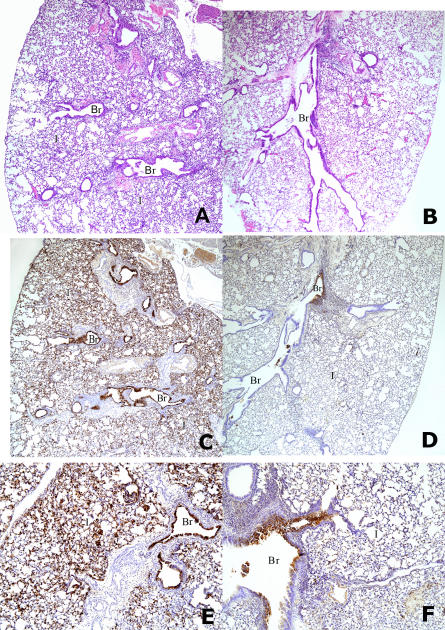
Figure 3. Histopathology in Pulmonary Tissue of Passively Immunized and Challenged Mice.
(A) Hematoxylin and eosin-stained lung sections (40×) revealed diffuse interstitial pneumonia (I) and bronchial and bronchiolar (Br) involvement in a mouse infected with influenza A/VN/1203/04 (H5N1) after i.p. injection of the control mAb D2.2.
(B) Mouse given mAb FLA5.10 prior to infection with influenza A/VN/1203/04 (H5N1), showing much less lung involvement than in (A).
(C) Immunohistochemistry (40×) revealed H5 antigen in bronchi, bronchioles (Br), and interstitial lesions (I) in a mouse given influenza A/VN/1203/04 (H5N1) following i.p. injection of the control mAb D2.2.
(D) H5 antigen in bronchus (Br) and not in bronchioles or interstitial areas (I) in a mouse given mAb FLA5.10 prior to influenza challenge.
(E) High magnification (100×) of (C) showing abundant H5 antigen in interstitial alveolar lesions (I) and bronchiolar epithelium (Br).
(F) High magnification of (D) (100×) showing H5 antigen only focally in a bronchus (Br) and not in the interstitial alveolar areas (I).

