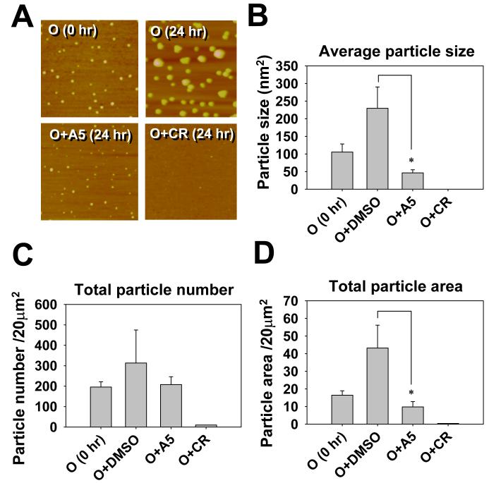
Fig. 5.
AFM data that demonstrate the ability of A5 to inhibit the growth of existing Aβ42 oligomers. Panel A shows representative 4 μm2 AFM images of Aβ42 oligomer particles. Pre-made Aβ42 oligomers were incubated at 25°C in the absence or presence of 10 equiv. of indicated compounds for 24 hr. DMSO was the solvent of the compounds and was added as the mock control in the sample labeled as O (24 hr). Pictures were taken from the 24-hr samples, except the one labeled as O (0 hr) was from the starting oligomer sample. The average particle size (B), the total particle number (C), and the total area covered by particles (D) were quantified by the Colony program (Fuji Photo Film, Japan). Data presented are mean ± standard error, n = 3, * p < 0.05.

