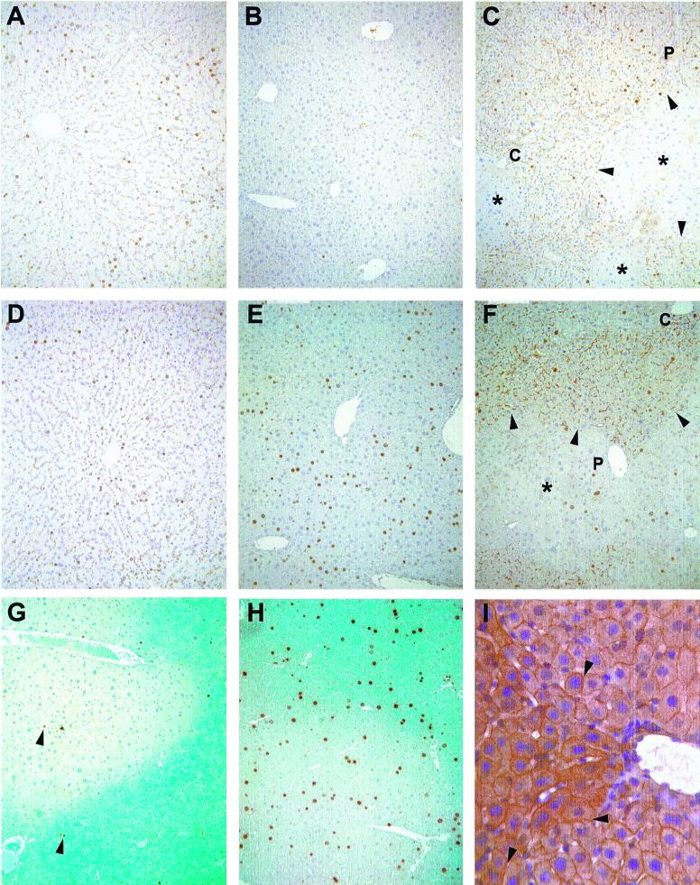
Figure 2.
Immunohistochemistry. Immunohistochemical detection of BrdUrd at 28 (A, B, C, and G) and 44 (D, E, F, and H) h posthepatectomy. (A and D) F344 rat. (B and E) nu/nu mouse. (C and F) MUP-uPA mouse repopulated with donor F344 rat hepatocytes. (G and H) MUP-uPA mouse repopulated with donor hPAP mouse hepatocytes. In C and F, arrowheads point to the edges of donor rat parenchyma, stained with mAb 106 to identify rat bile canilicular antigen (*, focus of endogenous mouse cells; P, portal tract; C, central vein). In G and H, blue staining indicates donor mouse parenchyma expressing the hPAP marker transgene. In G, arrowheads point to BrdUrd-positive hepatocyte nuclei. (A–G original magnification ×100.) (I) Immunohistochemical detection of connexin 32 in chimeric mouse liver. Arrowheads point to cytoplasmic junctions between mouse and rat hepatocytes that show connexin 32 staining (thin brown lines). The triangular patch of cells to the left of the vessel with slightly darker staining is composed of rat hepatocytes. Original magnification ×400.

