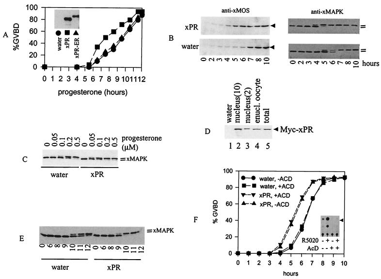
Figure 5.
xPR potentiates progesterone-induced MAP kinase activation and GVBD. (A) Oocytes (>60 oocytes per group) injected with water or xPR or xPR-ER mRNA were incubated with 0.5 μM of progesterone. GVBD was scored at the indicated time after the addition of progesterone. Shown is a representative of four independent experiments. (Inset) Typical expression of xPR or xPR-ER in mRNA-injected oocytes as determined via anti-Myc immunoblotting. We estimated that the amount of xPR derived from the injected mRNA was 5–10 times that of endogenous xPR, based on immunoblotting with anti-xPR antibodies (see Fig. 2C). (B) Oocytes injected with water (>200 oocytes) or xPR (>200 oocytes) were treated with progesterone (0.5 μM). At the indicated time, 20–25 oocytes were withdrawn randomly and lysed immediately. All samples were subjected to immunoblotting with anti-xMOS or anti-xMAPK, as indicated. Shown is a representative of three independent experiments. (C) Oocytes injected with water or xPR mRNA were incubated overnight with the indicated concentrations of progesterone and subjected to anti-xMAP kinase immunoblotting. Shown is a representative of three independent experiments. (D) Oocytes injected with water (lane 1) or Myc-xPR mRNA (lanes 2–5) were subjected to nuclear isolation. Extracts from intact oocytes (lanes 1 and 5, one oocyte each), enucleated oocytes (lane 4, one oocyte), or nuclei (lanes 2 and 3, 10 and two nuclei, respectively) were blotted with anti-Myc. Shown is a representative of two independent experiments. (E) Oocytes injected with water (100 oocytes) or xPR (100 oocytes) were individually enucleated (11). The nucleated oocytes were pooled before being divided into six groups of 15 each and treated with progesterone (0.5 μM). At the indicated time after the addition of progesterone, enucleated oocytes were lysed and subjected to immunoblotting with anti-xMAP kinase. Shown is a representative of three independent experiments. (F) Oocytes injected with water, or xPR mRNA, each were split into two groups (>60 oocytes per group) and immediately placed in OR2 or OR2 containing 5 μg/ml of AcD. After a 24-h incubation, progesterone (0.5 μM) was added to all four groups. GVBD was scored at the indicated time after the addition of progesterone. Shown is a representative of two independent experiments. (Inset) CAT assays of xPR-transfected COS cells treated with R5020 (1 μM), AcD (5 μg/ml), alone or in combination.

