Abstract
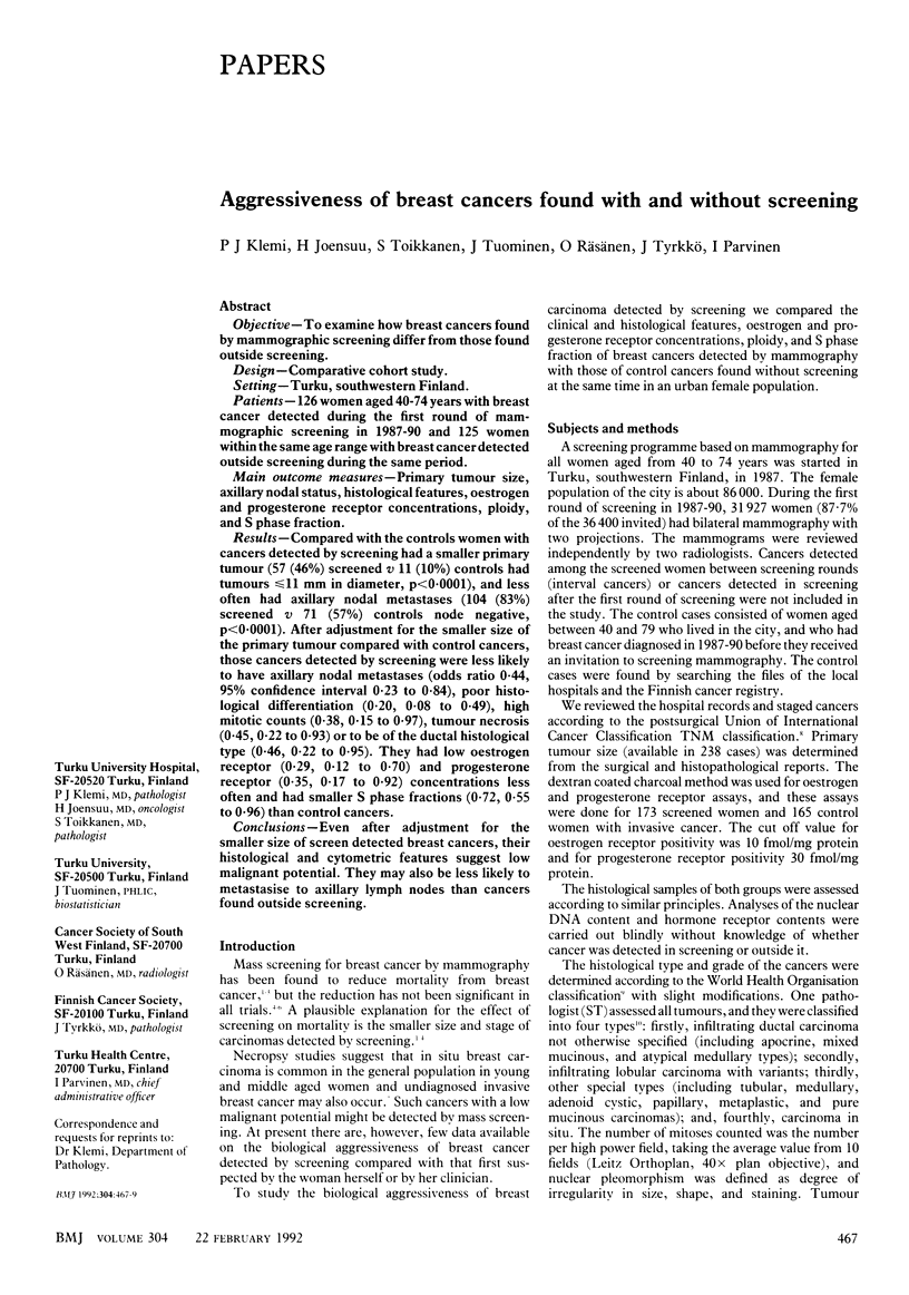
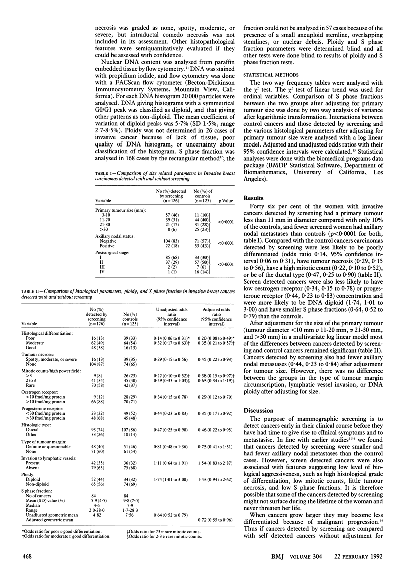
OBJECTIVE--To examine how breast cancers found by mammographic screening differ from those found outside screening. DESIGN--Comparative cohort study. SETTING--Turku, southwestern Finland. PATIENTS--126 women aged 40-74 years with breast cancer detected during the first round of mammographic screening in 1987-90 and 125 women within the same age range with breast cancer detected outside screening during the same period. MAIN OUTCOME MEASURES--Primary tumour size, axillary nodal status, histological features, oestrogen and progesterone receptor concentrations, ploidy, and S phase fraction. RESULTS--Compared with the controls women with cancers detected by screening had a smaller primary tumour (57 (46%) screened v 11 (10%) controls had tumours less than or equal to 11 mm in diameter, p less than 0.0001), and less often had axillary nodal metastases (104 (83%) screened v 71 (57%) controls node negative, p less than 0.0001). After adjustment for the smaller size of the primary tumour compared with control cancers, those cancers detected by screening were less likely to have axillary nodal metastases (odds ratio 0.44, 95% confidence interval 0.23 to 0.84), poor histological differentiation (0.20, 0.08 to 0.49), high mitotic counts (0.38, 0.15 to 0.97), tumour necrosis (0.45, 0.22 to 0.93) or to be of the ductal histological type (0.46, 0.22 to 0.95). They had low oestrogen receptor (0.29, 0.12 to 0.70) and progesterone receptor (0.35, 0.17 to 0.92) concentrations less often and had smaller S phase fractions (0.72, 0.55 to 0.96) than control cancers. CONCLUSIONS--Even after adjustment for the smaller size of screen detected breast cancers, their histological and cytometric features suggest low malignant potential. They may also be less likely to metastasise to axillary lymph nodes than cancers found outside screening.
Full text
PDF


Selected References
These references are in PubMed. This may not be the complete list of references from this article.
- Andersson I., Aspegren K., Janzon L., Landberg T., Lindholm K., Linell F., Ljungberg O., Ranstam J., Sigfússon B. Mammographic screening and mortality from breast cancer: the Malmö mammographic screening trial. BMJ. 1988 Oct 15;297(6654):943–948. doi: 10.1136/bmj.297.6654.943. [DOI] [PMC free article] [PubMed] [Google Scholar]
- Camplejohn R. S., Macartney J. C., Morris R. W. Measurement of S-phase fractions in lymphoid tissue comparing fresh versus paraffin-embedded tissue and 4',6'-diamidino-2 phenylindole dihydrochloride versus propidium iodide staining. Cytometry. 1989 Jul;10(4):410–416. doi: 10.1002/cyto.990100408. [DOI] [PubMed] [Google Scholar]
- Hedley D. W., Friedlander M. L., Taylor I. W., Rugg C. A., Musgrove E. A. Method for analysis of cellular DNA content of paraffin-embedded pathological material using flow cytometry. J Histochem Cytochem. 1983 Nov;31(11):1333–1335. doi: 10.1177/31.11.6619538. [DOI] [PubMed] [Google Scholar]
- Joensuu H., Toikkanen S., Klemi P. J. Histological features, DNA content and prognosis of breast carcinoma found incidentally or in screening. Br J Cancer. 1991 Sep;64(3):588–592. doi: 10.1038/bjc.1991.355. [DOI] [PMC free article] [PubMed] [Google Scholar]
- Kallioniemi O. P., Kärkkäinen A., Auvinen O., Mattila J., Koivula T., Hakama M. DNA flow cytometric analysis indicates that many breast cancers detected in the first round of mammographic screening have a low malignant potential. Int J Cancer. 1988 Nov 15;42(5):697–702. doi: 10.1002/ijc.2910420511. [DOI] [PubMed] [Google Scholar]
- Koscielny S., Tubiana M., Lê M. G., Valleron A. J., Mouriesse H., Contesso G., Sarrazin D. Breast cancer: relationship between the size of the primary tumour and the probability of metastatic dissemination. Br J Cancer. 1984 Jun;49(6):709–715. doi: 10.1038/bjc.1984.112. [DOI] [PMC free article] [PubMed] [Google Scholar]
- Nielsen M., Thomsen J. L., Primdahl S., Dyreborg U., Andersen J. A. Breast cancer and atypia among young and middle-aged women: a study of 110 medicolegal autopsies. Br J Cancer. 1987 Dec;56(6):814–819. doi: 10.1038/bjc.1987.296. [DOI] [PMC free article] [PubMed] [Google Scholar]
- Roberts M. M., Alexander F. E., Anderson T. J., Chetty U., Donnan P. T., Forrest P., Hepburn W., Huggins A., Kirkpatrick A. E., Lamb J. Edinburgh trial of screening for breast cancer: mortality at seven years. Lancet. 1990 Feb 3;335(8684):241–246. doi: 10.1016/0140-6736(90)90066-e. [DOI] [PubMed] [Google Scholar]
- Shapiro S., Venet W., Strax P., Venet L., Roeser R. Selection, follow-up, and analysis in the Health Insurance Plan Study: a randomized trial with breast cancer screening. Natl Cancer Inst Monogr. 1985 May;67:65–74. [PubMed] [Google Scholar]
- Tabár L., Fagerberg C. J., Gad A., Baldetorp L., Holmberg L. H., Gröntoft O., Ljungquist U., Lundström B., Månson J. C., Eklund G. Reduction in mortality from breast cancer after mass screening with mammography. Randomised trial from the Breast Cancer Screening Working Group of the Swedish National Board of Health and Welfare. Lancet. 1985 Apr 13;1(8433):829–832. doi: 10.1016/s0140-6736(85)92204-4. [DOI] [PubMed] [Google Scholar]
- Toikkanen S., Joensuu H. Prognostic factors and long-term survival in breast cancer in a defined urban population. APMIS. 1990 Nov;98(11):1005–1014. doi: 10.1111/j.1699-0463.1990.tb05027.x. [DOI] [PubMed] [Google Scholar]
- Verbeek A. L., Hendriks J. H., Holland R., Mravunac M., Sturmans F., Day N. E. Reduction of breast cancer mortality through mass screening with modern mammography. First results of the Nijmegen project, 1975-1981. Lancet. 1984 Jun 2;1(8388):1222–1224. doi: 10.1016/s0140-6736(84)91703-3. [DOI] [PubMed] [Google Scholar]


