Figure 5.
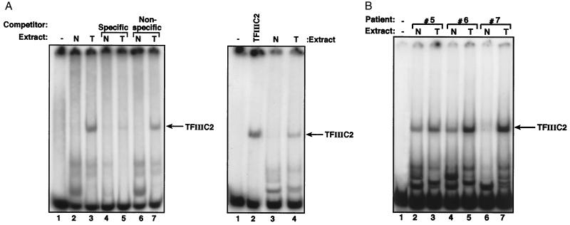
Ovarian tumors display elevated TFIIIC2 activity. (A Left) EMSA by using 1 ng of VA1 probe, 500 ng of polydI-dC, and no protein (lane 1) or 10 μg of protein extracts of normal ovarian tissue (N, lanes 2, 4 and 6) or ovarian tumor (T, lanes 3, 5 and 7) from patient 7. Lanes 4 and 5 also contain 60 ng of unlabeled VA1 gene fragment. Lanes 6 and 7 also contain 60 ng of unlabeled vector DNA of similar length. (Right) EMSA by using 1 ng of VA1 gene probe, 500 ng of polydI-dC competitor, and no protein (lane 1), 2 μg of partially purified TFIIIC2 (lane 2), or 10 μg of protein extracts of normal ovarian tissue (lane 3) or ovarian tumor (lane 4) from patient 7. (B) EMSA by using 1 ng of VA1 gene probe, 500 ng of polydI-dC competitor, and no protein (lane 1), or 10 μg of protein extracts of normal ovarian tissue (N, lanes 2, 4 and 6) or ovarian tumors (T, lanes 3, 5 and 7) from patients 5, 6, or 7, as indicated.

