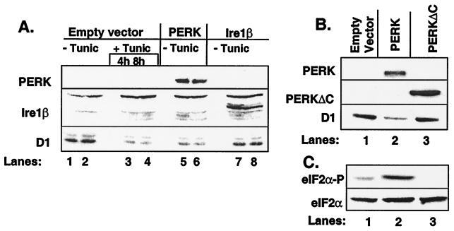
Figure 1.
PERK regulates accumulation of cyclin D1. (A) NIH 3T3 cells infected with empty virus (lanes 1–4), virus encoding PERK (lanes 5–6), or virus encoding Ire1β (lanes 7–8) were left untreated or were treated with 0.5 μg/ml tunicamycin (lanes 3–4). Equivalent amounts of cellular protein were resolved on denaturing polyacrylamide gels and subjected to immunoblot analysis with the 9E10 antibody, which recognizes a c-myc epitope tag expressed at the C terminus of both PERK and Ire1β or a cyclin D1-specific monoclonal antibody. Sites of antibody binding were visualized by enhanced chemiluminescence. (B and C) After infection of NIH 3T3 cells with virus encoding PERK (lane 2), PERKΔC (lane 3) or empty virus (lane 1), equivalent amounts of total protein were resolved on a denaturing polyacrylamide gel and transferred to nitrocellulose membrane. Membranes were subsequently probed with the 9E10 antibody (PERK and PERKΔC) or antibodies specific for cyclin D1, serine 51-phosphorylated eIF2α, or total eIF2α. Sites of antibody binding were visualized by enhanced chemiluminescence.

