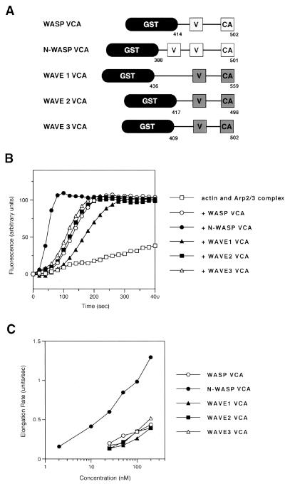
Figure 1.
Comparison of Arp2/3 complex-mediated actin polymerization activity of WASP family VCAs. (A) Schematic structures of WASP family VCAs. VCA regions of WASP family proteins were constructed as GST fusion proteins. The numbers shown in the figure refer to those of amino acid residues in the full-length protein. V, verprolin homology domain; CA, cofilin homology domain and acidic region. (B) Actin polymerization was followed in the presence of 100 nM VCAs using pyrene actin. Fluorescence intensity is given in arbitrary units. (C) Dose-response curve showing the change in the maximum rate of filament elongation as a function of increasing concentrations of VCAs.

