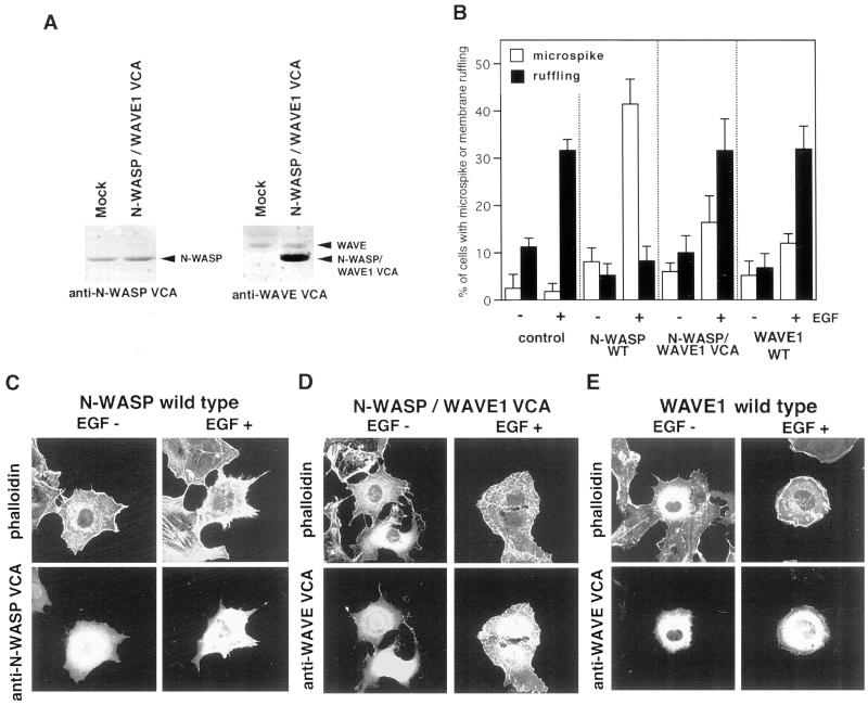
Figure 4.
N-WASP VCA is necessary for actin–microspike formation induced by N-WASP. (A) Confirmation of chimerization. Cell lysates of COS7 cells overexpressing wild-type N-WASP and N-WASP/WAVE1 VCA were subjected to Western blotting with anti-N-WASP VCA and anti-WAVE1 VCA antibodies. (B) Quantitation of microspike and membrane ruffle formation. Transfected cells were serum-starved and then stimulated with or without EGF for 10 min. The percentage of cells forming microspikes or membrane ruffles was calculated among transfected cells. Error bars represent the standard deviation of three different determinations. At least 50 cells were counted in each determination. (C–E) Immunofluorescence staining of transfected cells. Cells were stained with phalloidin to visualize actin filaments and antibody against N-WASP VCA or WAVE1 VCA to detect ectopically expressed proteins.

