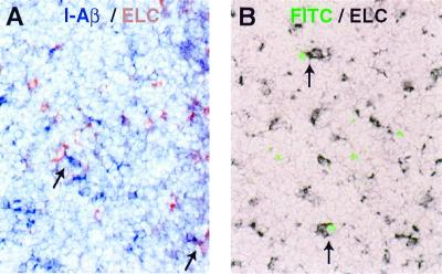
Figure 6.
Close proximity of ELC-producing cells and DCs. (A) Double in situ hybridization of LN for I-Aβ (blue) and ELC (red). (B) Section from the draining LN of a FITC skin-painted mouse, taken at day 1 after painting. Newly immigrated DCs are identified by strong intracellular FITC fluorescence (green) and ELC mRNA-expressing cells by in situ hybridization (black). Arrows show examples of colocalizing cells. (Magnification, ×20.)

