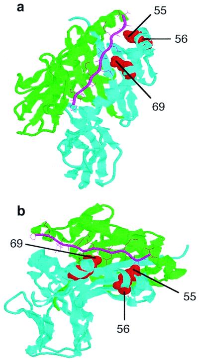
Figure 6.

Modeling of the HLA-DP molecule based on the DR1 molecule. (a) Top view of the DR1 peptide-binding cleft, with a green α-chain and a cyan β-chain. (b) Side view of the DR1 peptide binding groove. The HA306–318 peptide is indicated by pink sticks. The polymorphic charged residues at positions 55–56 and 69, indicated as red balls, are located in the peptide binding region. Despite the appearance from the ribbon structure, minimal, if any, portions of these polymorphic residues are exposed on the outside or top of the β-chain.
