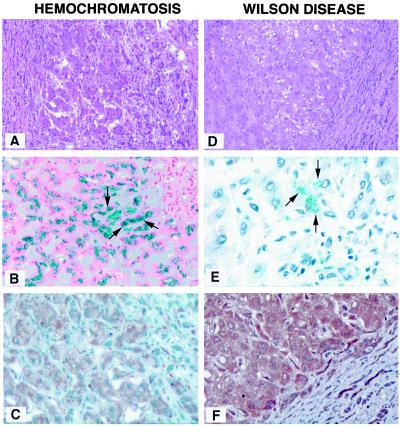
Figure 2.
Sections from patients with hemochromatosis (A–C) or WD (D–F). (A) Hematoxylin/eosin stain, which shows hyperplastic nodule. Surrounding the nodule, regenerative fibrous tissues and mononuclear inflammatory cells are present, indicating the presence of cirrhosis (×100). (B) The iron stain (Prussian blue), which displays iron granules in hepatocytes (arrowheads, ×400). The NOS2 expression is seen predominantly in the cytoplasm of hepatocytes. (C) In addition, NOS2 also is overexpressed in endothelial cells within areas of bridging fibrosis. The H&E stain in WD (D) shows regenerative and hyperplastic nodule with fibrosis and inflammation, indicating the presence of cirrhosis (100×). Confirmatory rhodamine stain shows the intracellular deposition of copper (E, ×400). The immunohistochemical stain with anti-NOS2 is shown (F). NOS2 overexpression is seen within hepatocytes of hyperplastic nodule (×200).

