
Figure 2.
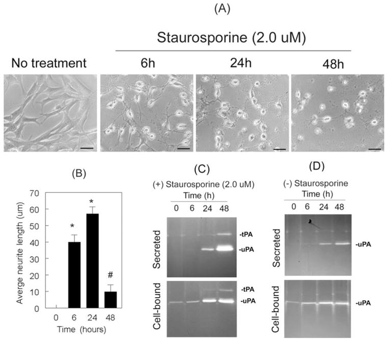
Differentiation of RGC-5 cells was associated with the expression of tPA and uPA. RGC-5 cells (4 × 10³ cells/mL) were left untreated or were treated with 2.0 μM staurosporine (in serum-free medium). At 6 hours, 24 hours, and 48 hours after treatment, their morphology was observed under a phase-contrast microscope. Conditioned medium (CM) was collected from untreated and treated cells, and proteolytic activities of tPA and uPA were determined by zymography assays. (A) Staurosporine induced the differentiation of RGC-5 cells in a time-related fashion and (B) led to an increase in neurite length at 6 hours and 24 hours (scale bar, 40 μm). At 48 hours, however, RGC-5 cells had reduced neurite length. *P < 0.05, compared with untreated cells. #P < 0.05, compared with cells treated for 24 hours. (C) At 6 hours, zymography assays did not detect proteolytic activity of uPA or tPA, though RGC-5 cells had already differentiated. Differentiated RGC-5 cells synthesized uPA (C, lower panel) and secreted it into the conditioned medium at 24 hours (C, upper panel). At 48 hours they also expressed tPA. Undifferentiated RGC-5 cells did not express tPA, but they expressed uPA.