
Figure 3.
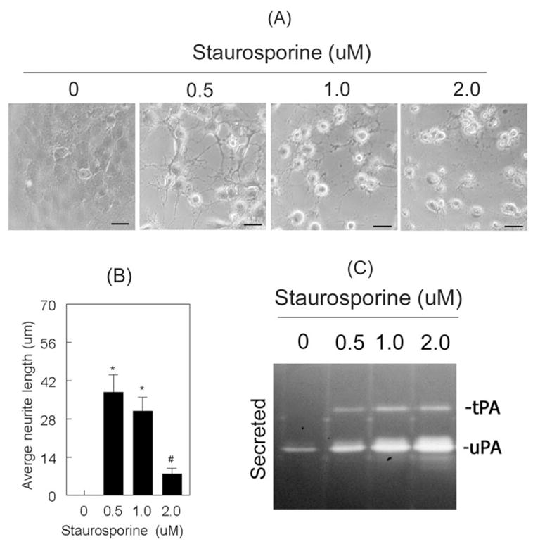
Staurosporine induced proteolytic activities of tPA and uPA in a dose-dependent fashion. RGC-5 cells (4 × 10³ cells/mL) were left untreated or were treated for 48 hours with 0.5 μM, 1.0 μM, and 2.0 μM staurosporine (in serum-free medium). (A) RGC-5 cells treated with staurosporine (up to 1.0 μM) differentiated and had increased neurite outgrowth. However, at a concentration of 2.0 μM, neurite outgrowth was significantly reduced (B). Scale bar, 40 μm. *P < 0.05, compared with untreated cells. #P < 0.05, compared with 0.5 μM staurosporine-treated cells. (C) RGC-5 cells left untreated expressed low levels of uPA but not tPA, whereas cells treated with staurosporine expressed increased levels of both tPA and uPA.