
Figure 4.
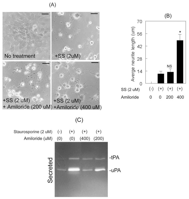
Inhibition of uPA proteolytic activity increased neurite outgrowth. RGC-5 cells (4 × 10³ cells/mL) were left untreated or were treated for 48 hours with 2.0 μM staurosporine (SS) or with 2.0 μM staurosporine (SS) and indicated concentrations of amiloride. (A, B) Compared with untreated cells, staurosporine-treated cells differentiated but neurites were shorter. RGC-5 cells treated with 2.0 μM staurosporine and 400 μM amiloride also differentiated, but these cells exhibited increased neurite length than did cells treated with 2.0 μM staurosporine alone or cells treated with staurosporine and 200 μM amiloride. Scale bar, 40 μm. *P × 0.05, compared with cells treated with staurosporine alone (NS). (C) CM collected from untreated cells indicated low levels of uPA, whereas CM collected from staurosporine-treated cells showed increased levels of tPA and uPA. In contrast, CM collected from cells treated with 400 μM amiloride along with staurosporine showed a reduction in the proteolytic activity of uPA.