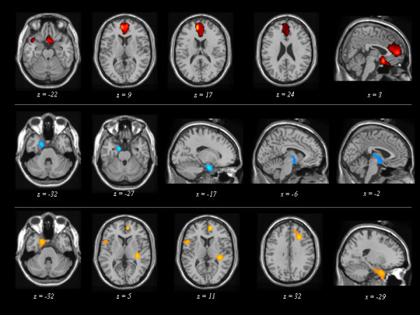
Figure 3.

Maps of significant voxels representing regions of hypoperfusion in FTLD patients according to the three Latent Classes, superimposed to reference T1-weighted MRI image. LC: Latent Class. LC1 (first row), LC2 (second row), and LC3 (third row) patient subgroups are reported. Statistical threshold, P < 0.001, T ≥ 3.40, minimum cluster size = 50 voxels. Neurological convention: left is on the left side and vice versa.
