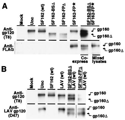
Figure 4.
Mixed oligomer formation detected by coimmunoprecipitation of Env variants. Effector cells prepared as in Fig. 2 were lysed in 1% Nonidet P-40, and the lysates were divided into two tubes. HIV-1 Env was immunoprecipitated as designated on the left with either the broadly cross-reactive T8 anti-gp120 mAb, the anti-FLAG epitope tag mAb, or the D47 anti-gp120 mAb that specifically recognizes the V3 loop of LAV Env but not SF162. As a control (mixed lysates), lysates from cells separately transfected with either SF162-BSΔ or SF162-FP* alone were mixed before immunoprecipitation. Immunoprecipitates were analyzed by SDS/PAGE and immunoblotting with the broadly cross-reactive T8 anti-gp120 mAb, as described in Materials and Methods. Another control involved treatment under the identical transfection conditions without DNA (Mock). The symbols Δ and * indicate truncated and FLAG-tagged Envs, respectively. The positions of full-length gp160 (gp160) and truncated gp160 (gp160Δ) are indicated on the right.

