Figure 4.
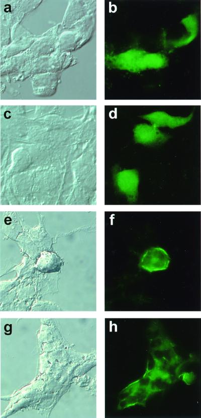
25-Dx is localized to the cell membrane of hypothalamic neurons. GT1–7 cells, grown on glass coverslips, were transfected with control GFP expression plasmid pEGFP-N1 (CLONTECH) (a–d) or the GFP/25-Dx fusion protein expression plasmid pEGFP/25-Dx (e–h). Forty-eight hours after transfection, the cells were fixed and mounted on microscope slides and photographed using bright-field microscopy enhanced with Nomarski optics (40× magnification) (Left). To the right of each bright-field image is the same visual field photographed under UV illumination, using a filter set designed for FITC fluorescence. The GFP protein fluoresces uniformly green throughout the cytoplasm (b and d), whereas the GFP/25-Dx fusion protein exhibits more intense green fluorescence at the cell border (f and h).

