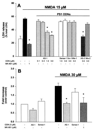
Figure 5.
Effects of PS1 ODN treatment on NMDA-induced neurotoxicity (A) and PS1 mRNA expression (B) in primary cortical neurons from wt mice. (A) Cortical neurons were exposed to increasing concentrations of different ODNs, ranging from 0.1 μM to 3 μM, for 2 h before and during the NMDA treatment. Cell death was quantified by measuring the release of LDH enzyme and was expressed as the percentage of total LDH release elicited by 100 μM NMDA. (B) Primary cortical neurons were exposed to different ODNs for 2 h before the NMDA treatment. PS1 mRNA levels were measured by Northern blot analysis 1 h after NMDA. Data represent mean ± SEM of at least three different experiments and are from three separate cell preparations. MK-801 was added to the culture media 10 min before NMDA. ODN sequence and acronimus are reported in Experimental Procedures. *, P < 0.01 vs. NMDA alone treated values.

