
Figure 1.

Increased osmolarities permit less dye release. (A)
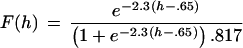
Percent of FM1–43 that escapes per quantum of transmitter released by
hypertonic stimulation, relative to dye released per quantum with
action potentials, as a function of hypertonicity. Figure presents data
from 780 synapses in 8 experiments on conventional and microdot
cultures; error bars indicate one SEM. The smooth line is given by
where h is the hypertonicity in
osmolars. See text for discussion of equation. Dye release was
determined for hypertonic and action potential stimulation in the same
synapses. (B) Image of field after loading as described in
text (Left) and after prolonged stimulation to release all
dye in synaptic vesicles (Right). Synapses appear as small
black dots (Left) that are no longer visible
(Right).
 |