
Figure 5. Conformation of the ssDNA in the closed, intermediate, and closed forms.
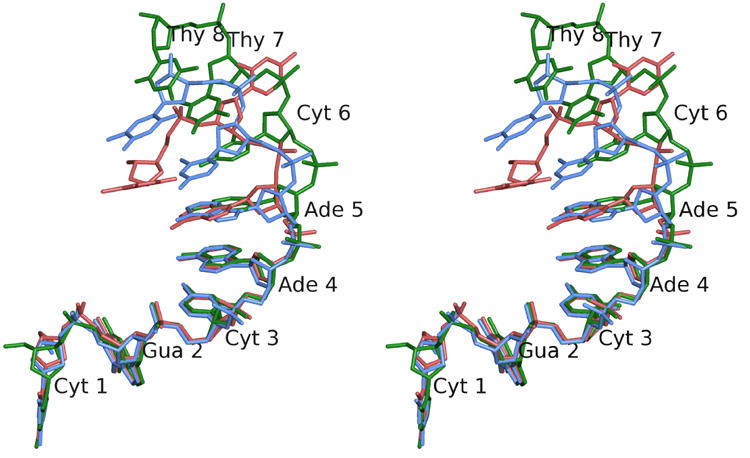
Stereo diagram of the ssDNA in the closed (red), intermediate (blue), and open (green) conformations. The diagram illustrates the differences in conformation of the ssDNA as it enters the active site. The first five nucleotides remain essentially unchanged, while nucleotides 6 and 7 change conformation to approach the active site. Nucleotide 8 is ordered only in the open conformation. Note the base of Thy 7, which is in a completely different conformation in the closed form. As the structure changes from the closed to the open forms, the phosphate of Thy 7, the scissile phosphate, approaches the active site. Nucleotides are numbered from the 5′ to the 3′ end starting at Cyt 1.