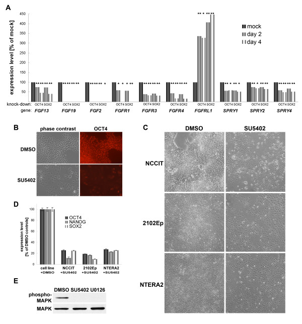
Figure 3.
Autocrine FGF signaling is cruicial for hEC cell self-renewal. (A): Expression changes of genes involved in FGF signaling. Samples were assayed 2 and 4 days after transfection. Transcripts of FGF19, FGF2, and FGFR1 (at 96 h) were undetectable in the OCT4 and SOX2 RNAi samples. (B): Immunostain of OCT4 protein in NCCIT hEC cells 5 days after SU5402 treatment indicating loss of self-renewal. (C): Cellular morphology after 5 days of SU5402 treatment vs. DMSO controls using three different hEC cell lines. (D): Monitoring of OCT4, NANOG, and SOX2 expression levels by real-time PCR in samples from (C). Error bars indicate technical variation (DMSO control: means between cell lines). (E): Short-term effect of SU5402 treatment on MAPK phosphorylation in NCCIT hEC cells. Total MAPK protein (bottom panel) served as loading control. U0126 is a specific inhibitor of MEK (MAPKK) which directly phosphorylates ERK (MAPK). This sample served as positive control for the assay.

