
Figure 3.
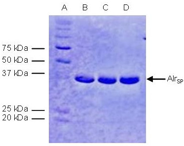
SDS-polyacrylamide gel electrophoresis of alanine racemase from S. pneumoniae (Lanes B-D) and protein molecular weight markers (Lane A, BioRad Dual Color Marker) stained with Coomassie blue. Lanes B-D represent the three peak fractions from the final gel filtration step.