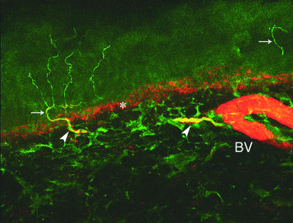
Fig 5 Skin biopsy taken at the distal leg in a healthy subject. Double staining confocal microscopy with antibodies against protein gene product 9.5 (green), which stain nerve fibres, and antibodies against collagen IV (red), which stain the dermal-epidermal junction (asterisk) and blood vessels (BV). Arrows indicate intraepidermal nerve fibres and arrowheads indicate dermal nerve bundles. Note the branched intraepidermal fibre arising from a dermal nerve fibre (original magnification ×40)

An official website of the United States government
Here's how you know
Official websites use .gov
A
.gov website belongs to an official
government organization in the United States.
Secure .gov websites use HTTPS
A lock (
) or https:// means you've safely
connected to the .gov website. Share sensitive
information only on official, secure websites.
