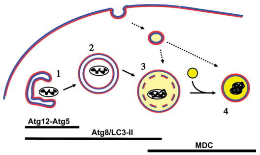
FIGURE 1.

Schematic of AV maturation. Pre-autophagosomal membranes may be derived from smooth endoplasmic reticulum. Conjugation of Atg12-Atg5 complex and of Atg8/LC3 to the membrane is required for extension of the sequestering membrane (1). In contrast to other organelles such as endoplasmic reticulum, Golgi, or endosomes in which the lumen contacts ‘‘extracellular’’-type membrane (blue), the autophagosome (2) is a double membrane structure with cytoplasmic leaflets both inside and out (red). Upon completion of the nascent autophagosome, Atg12-Atg5 dissociates, while a portion of LC3-II remains associated with the AV until degraded. Early acidification (light yellow) and dissolution of the inner membrane (3) occurs, most likely through fusion with the endocytic pathway, and appears to be necessary for fusion with lysosomes (bright yellow) to form fully degradative autolysosomes (4). Acidification and membrane compaction results in staining of autolysosomes by MDC. Markers of late AVs are useful for measuring autophagic stress, but are less specific as convergence with endosomes and other vesicular pathways represent a normal consequence of AV maturation.
