
Figure 5.
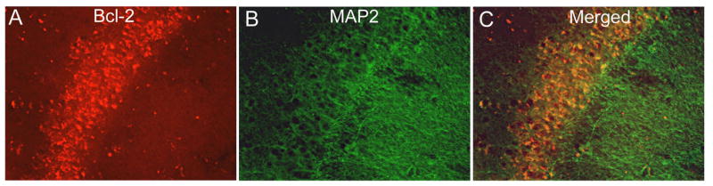
Colocalization of bcl-2 with neurons in APP mice. APP mouse brain sections were immunofluorescently labeled to reveal bcl-2 (red) in the molecular layer of the hippocampus (Panel A). Panel B shows MAP2 (green) labeled neurons in the pyramidal layer of the hippocampus. Panel C shows the micrographs merged to show colocalization of Bcl-2 in the cytoplasm of neurons (yellow regions represent colocalization). Micrographs were taken at 400× final magnification.