Figure 1.

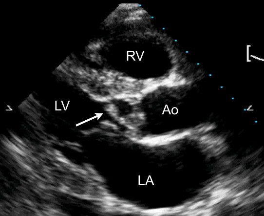
A transthoracic parasternal long axis view with harmonic imaging illustrating a vegetation (arrow) involving the aortic valve during diastole. LA, left atrium; LV, left ventricle; RV, right ventricle; Ao, aorta.

A transthoracic parasternal long axis view with harmonic imaging illustrating a vegetation (arrow) involving the aortic valve during diastole. LA, left atrium; LV, left ventricle; RV, right ventricle; Ao, aorta.