
Fig. 3.
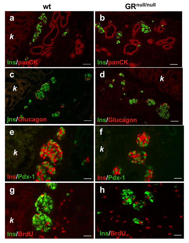
Rescue of endocrine differentiation upon grafting the GRnull/null pancreas. Three pancreata from GRnull/null (b, d, f, h) or GR+/+ littermates (a, c, e, g) fetuses at E15.5 were grafted for 7 days under the kidney capsule of SCID mice. One hour before killing, the animals were injected with BrdU, as described in Material and methods. Both groups of grafted tissue showed indistinguishable immunohistochemical patterns: ducts expressing pan-cytokeratin (panCK) (red, a, b) were intermingled with endocrine cell clusters (green, a, b). Islets were composed of glucagon-expressing cells (red, c, d) surrounding a core of insulin (Ins)-positive cells (green, c, d). All the insulin-positive cells (red, e, f) had matured and expressed Pdx-1 (green nuclei, e, f). Beta-cell proliferation rates analysed by double immunohistochemistry for BrdU (red nuclei) and insulin (green) were identical in both groups of grafted tissue (g, h). k, kidney; wt, wild-type. Scale bars=50 μm (a–d) and 25 μm (e–h)