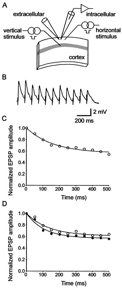
Figure 1.
Short-term synaptic dynamics in the thalamocortical slice. (A) Schematic diagram of the position of the recording and stimulating electrodes positions in primary somatosensory cortex. The gray band represents layer 4 of the barrel field. A field electrode was used to apply bicuculline methiodide focally. (B) Average responses (3 trials) evoked by stimulation of the horizontal layer 2–2 pathway at 20 Hz in one slice from deprived cortex. (C) Single exponential fit to the responses in B. (D) Single exponential fit to the responses evoked by 20-Hz stimulus trains averaged across all recordings in deprived cortex (open circles, n = 14 neurons from 10 slices) and spared cortex (filled circles, n = 12 neurons from 10 slices) of rats trimmed for 5 days.

