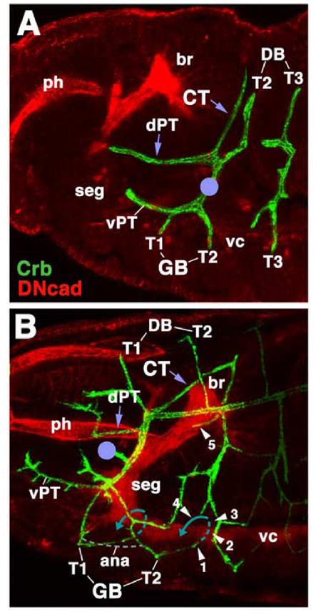
Fig.2.

Embryonic origin of the cerebral trachea and ganglionic tracheal branches. Both panels show Z-projections of confocal sections of embryos (lateral view, anteriior to the left) labeled with anti-Crb (green) to visualize tracheae. Anti-DN-cadherin (red) labels neuropile and other embryonic structures. A: Stage 14. Cerebral trachea (CT) and dorsal pharyngeal trachea (dPT) form a Y-shaped, anteriorly directed branch of the first segmental trachea (I) that grow around the posterior surface of the brain (br). Other branches of the first segmental trachea are the dorsal branches (DB) of segments T2 and T1 (formed later than stage 14), the ventral ganglionic branches (GB) of segments T1 and T2, and the ventral pharyngeal trachea. The location of the anterior spiracle is indicated by violet circle. B: Stage 15 late. Segmental tracheae have fused, primary branches have increased in length, and some secondary branches have been initiated. Note position of the cerebral trachea (CT) and dorsal pharyngeal trachea (dPT). The cerebral trachea has reached the medial surface of the brain neuropile (arrowhead ‘5’). Ventral ganglionic branches contact the ventral surface of the neuropile of the ventral nerve cord (vc; arrowhead ‘1). During later stages ganglionic branches will extend underneath the neuropile, turn dorsally (hatched blue line; arrowheads ‘2’ and ‘3’) and then laterally (solid blue line, arrowhead ‘4’). Anastomoses (ana; gray hatched line) will interconnect ganglionic branches of neighboring segments.
Other abbreviations: ph pharynx; seg subesophageal ganglion
