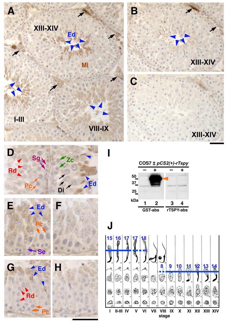
Figure 2.

Detection of spermatid-specific expression of rat Tspy using immunohistochemistry. (A) Only elongating spermatids at various spermatogenic stages were stained positive for the anti-rTspy antiserum (arrowheads). Roman numbers indicate spermatogenic stages as described in J. (B) Image of an adjacent section immunostained similarly by a control antiserum preabsorbed with GST-rTspy. The staining of elongating spermatids was significantly reduced (arrowheads). (C) Image of an adjacent section immunostained by a preimmune serum as a negative control. (D) Elongating spermatids at stage XIII are positively stained (blue arrowheads), but spermatids of stage II–III were negative (red arrowheads). No other cells, including somatic cells, were positive for the antiserum (Sg, Zc, Pc and Di). (E–H) Sections of seminiferous tubules at spermatogenic stage VIII (E and F) and II–IV (G and H) immunostained by the anti-rTspy antiserum (E and G), and control antiserum preabsorbed with GST-rTspy (F and H). Only elongating seprmatids are faintly positive (blue arrowheads). Abbreviations; Di, diplotene spermatocytes; Ed, elongating spermatids; Mi, meiotic dividing cells; Pc, pachytene spermatocytes; Rd, round spermatids; Se, Sertoli cells; Sg, spermatogonia; Zc, zygotene spermatocytes. Scale bars = 50 μm in A–C and D–H, respectively. (I) Western-blot of COS7 cells transfected with a rat Tspy expression vector using the anti-rTspy antiserum pre-absorbed with GST alone (lanes 1 and 2; GST-abs) and with GST-rTspy fusion protein (lanes 3 and 4; rTspy-abs). Positive band for rat Tspy protein was detected in COS7 cells transfected with rat Tspy expression vector (lane 2), but not in non-transfected cells (lanes 1, 3) or those reacted with the same antiserum subtracted with GST-rTspy protein (lane 3, 4). Anti-rTspy serum detected recombinant rTspy (lane 2). (J) Spermatogenic stages of the rat testis. Blue lines indicate the immuno-positive cells in steps 8–18 of spermatid development. Romanic numbers indicate stages of spermatogenesis. Dotted lines represent sections showing faint staining of spermatids in these stages. Modified from review by Russell et al. [28].
