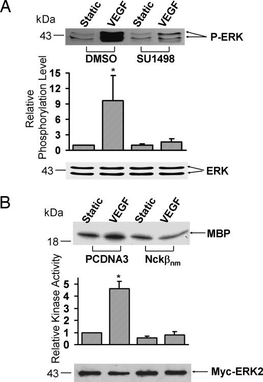
Fig. 2.
Flk-1 and Nckβ mediate the VEGF-induced ERK activation. (A) BAECs were treated with 0.1% DMSO (the solvent of SU1498) as control or 5 μM SU1498 for 1 h. (B) Myc-ERK2 was cotransfected with control vector PCDNA3 or Flag-Nckβnm into BAECs. These cells were then subjected to VEGF (10 ng/ml) for 15 min or kept under static incubation. (A) The upper gel band shows the phosphorylated ERK levels, and the lower gel band represents the loaded ERK proteins under different conditions as indicated. (B) The cell lysates were immunoprecipitated with anti-Myc antibodies for immunocomplex kinase assays by using myelin basic protein (MBP) as the substrate. The upper gel band is phosphorylated MBP, which indicates the level of ERK activation, and the lower gel band shows IB with an anti-Myc antibody to indicate that the levels of the expressed exogenous Myc-tagged ERK proteins were comparable among the various samples. The bar graphs are the results of densitometry analysis showing mean ± SEM from three separate experiments. The asterisks indicate significant differences (P < 0.05) between the SU1498-treated sample and DMSO control after VEGF activation in A and between cells transfected with PCDNA3 and Nckβnm after VEGF activation in B.

