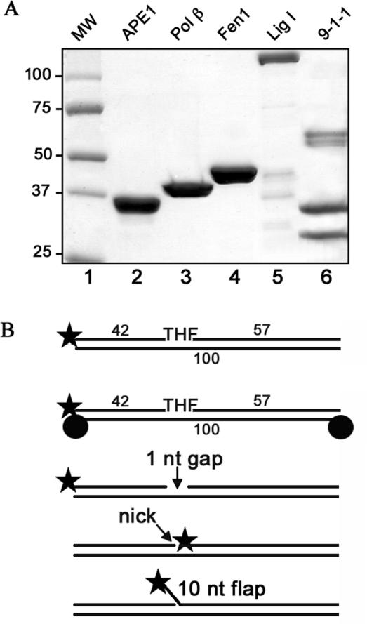
Figure 1.
Proteins and substrates used in reconstitution of long patch base excision repair in vitro. (A) Recombinant proteins were purified as described in ‘Materials and Methods’ and separated on a 8–20% gradient SDS-PAGE gel, and stained with Coomassie Blue. Lane 1: molecular weight markers; lane 2: APE 1 (2 μg); lane 3: Pol β (2 μg); lane 4: Fen 1 (2 μg); lane 5: Lig I (2 μg); lane 6: 9-1-1 complex (6 μg). (B) Schematic representation of the 32P-5′-labeled oligonucleotide substrates used in the study: a 100 bp duplex oligonucleotide containing a THF moiety at the position 43 was used for repair reactions, the ends of the substrate were either free (unblocked substrate) or blocked with a biotin at each end (blocked substrate); a 100 bp duplex oligonucleotide with a 1 nucleotide gap at the same position was used for the Pol β assay; with a nick for the Lig I assay and with a 10 nucleotide flap for the Fen 1 reaction

