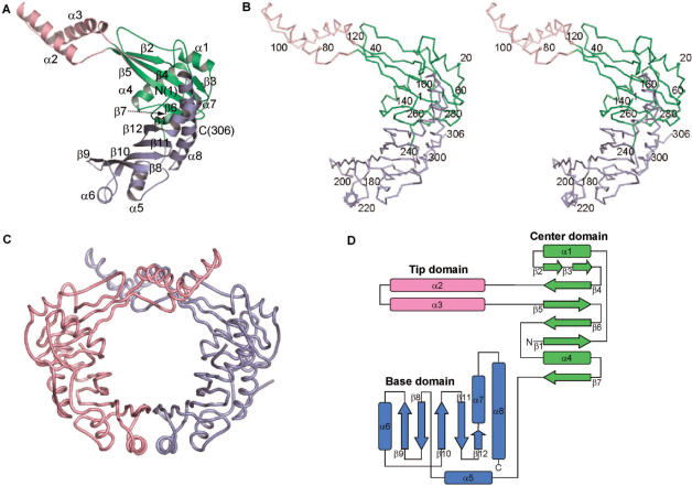
Figure 1.
Overall structure of P. aeruginosa RdgC. (A) Ribbon diagram of a monomer. Secondary structure elements were assigned by the TOPS server (http://tops.ebi.ac.uk/tops/). (B) Stereo Cα trace of a monomer. Every tenth residue is marked by a dot and every twentieth residue is labeled. (C) Pseudomonas aeruginosa RdgC dimer. A and B chains are colored in light purple and pink, respectively. (D) Topology diagram. β-Strands are depicted as arrows and α-helices as rectangles. All figures except Figures 1D, 3 and 4D are drawn with PyMOL (DeLano, 2002, The PyMOL Molecular Graphics System, http://www.pymol.org/).

