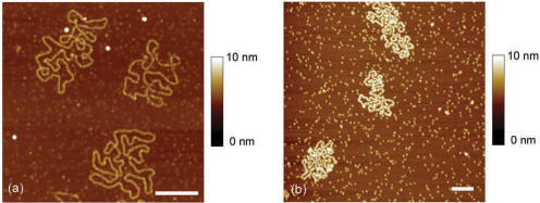
Figure 4.
(a) AFM image of M13 ssDNA–gp32 complexes formed in spermidine buffer Tris 20 mM pH 7.5, NaCl 300 mM, SpdCl3 300 µM with R = [(gp32)/(nucleotides)] = 1/7. (b) AFM image of M13 ssDNA–yRPA complexes formed in spermidine buffer Tris 20 mM pH 7.5, NaCl 20 mM, SpdCl3 50 µM with R = [(yRPA)/(nucleotides)] = 1/10 (scale bars 500 nm).

