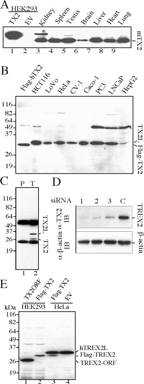
Figure 2.
Expression of endogenous TREX2 in mouse tissues and human cell lines. (A) Mouse tissues. Lanes 1 and 2: lysates from human HEK293 cells transiently transfected with TX2 (pCMV-Flag-TREX2) or EV (pCMV empty vector). Flag-TREX2 is the fusion product of 19 amino acids (including Flag-peptide) fused with the 26-kDa human TREX2 ORF. The predicated molecular weight is 28 kDa. (B) Human cell lines. Flag-TREX2: lysate from pCMV-Flag-TREX2-transfected HEK293 cells. (C) Immunoprecipitation-western blot using HeLa cells. Immunoprecipitation complex by either pre-immune serum (lane 1) or anti-TREX2 antibodies (lane 2) was resolved by SDS-PAGE, followed by anti-TREX2 western blot. (D) Endogenous TREX2–siRNA knockdown. HeLa cells transfected with each of TREX2–siRNA duplexes (lanes 1, 2 and 3, Table 1) or control siRNA (lane C) were collected 72 h post-transfection. Cell lysates were prepared and subjected to western blot with anti-TREX2 antibody. β-Actin serves as a loading control. (E) Examination for post-translational modification of TREX2. Lane 1: 26-kDa TREX2 open-reading frame (ORF) (25) expressed in HEK293 cells. Lane 2: Flag-tagged 26-kDa TREX2 expressed in HEK293 cells. Lane 3: Flag-tagged 26 kDa TREX2 expressed in HeLa cells. Lane 4: empty vector-transfected HeLa cells.

