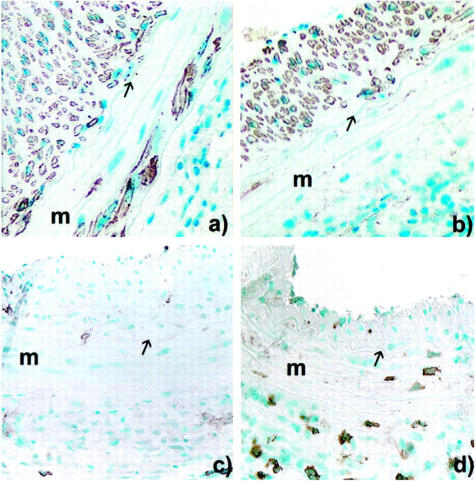
Figure 6.

Peroxidase immunocytochemistry of allografted aortic segment at 60 days after a long allogeneic exposure in: CD8+ T cell-depleted animals (a) and wild-type animals (b) using anti-α-actin mAb (HHF35); and CD8+ depleted-animals (c) and wild-type animals (d) stained using anti-CD8 mAb (MRC-OX-8). Original magnification, ×200. m, Media, arrow, internal elastic lamina.
