
Figure 5.
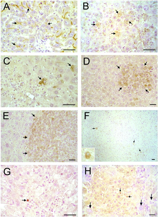
Immunohistochemical detection of hepatocyte markers in livers of retrorsine-exposed rats after PH. A and B: Indirect immunoperoxidase analysis of Pgp on liver sections from retrorsine-exposed rats 5 and 3 days after PH, respectively. C–F: Indirect immunoperoxidase analysis of AFP on liver sections from retrorsine-exposed rats 1, 5, 14, and 23 days after PH, respectively. G and H: Indirect immunoperoxidase analysis of WT1 on liver sections from retrorsine-exposed rat 1 and 5 days after PH. In hepatocytes and SHPCs 5 days after PH (A), Pgp is localized to the bile canaliculi. However, a subset of SHPCs express cytoplasmic Pgp 3 days after PH (B). Small, single AFP-positive hepatocyte-like cells in retrorsine-exposed rat livers are present 1 day after PH (C). Rapidly proliferating SHPCs 5 days after PH form AFP-positive clusters (D). Most SHPCs continue to strongly express AFP through 14 days after PH (E). By 23 days after PH, most SHPCs have ceased expression of AFP, and few AFP-positive cells remain (F). Rare growth-arrested megalocytes are weakly to moderately AFP-positive through 5 days after PH, after which they express trace amounts of AFP at levels similar to those of control rats (the AFP antibody weakly cross-reacts with the adult form of AFP). WT1 protein is localized to the nucleus of rare liver parenchymal cells in retrorsine exposed rats 1 day after PH (G) and is weakly expressed 5 days after PH by proliferating SHPCs and is not expressed by fully differentiated hepatic megalocytes (designated by long arrows) (H). WT1 protein is localized to the nucleus of a small fraction (approximately 5%) of proliferating SHPCs (small arrows, H). Bar, 50 μm. Small arrows, small hepatocyte-like cells. Large arrows (H), growth-arrested, fully differentiated hepatocytes. Note: The inset of F shows a higher magnification of a representative AFP-positive hepatocyte from a different tissue section 23 days after PH.