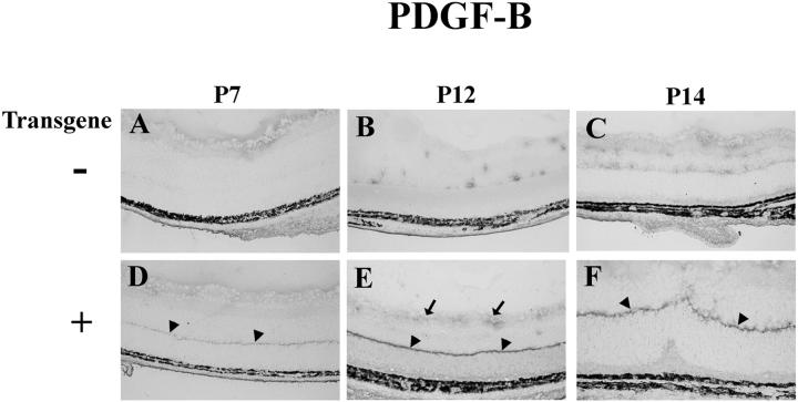
Figure 3.
Immunohistochemical staining for PDGF-B in the retinas of transgene-negative (−) or -positive (+) rho/PDGFB1 mice. Retinal frozen sections were immunohistochemically stained for PDGF-B as described in Materials and Methods. All mice show some diffuse staining on the surface of the retina at P7. At P12 and P14 control mice show focal staining around blood vessels. Transgene-positive mice show a discrete band of staining at the inner border of the outer nuclear layer (arrowheads), the region of photoreceptor terminals. The band is faint at P7 and more intense at P12 and P14. There is also more staining at the surface of the retina in transgenics at P12.

