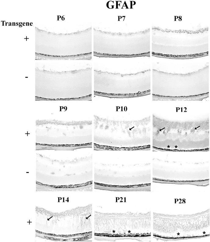
Figure 5.

Immunohistochemical staining for glial fibrillary acidic protein (GFAP) in transgene-positive (+) and -negative (−) rho/PDGFB1 mice. There is no difference in GFAP staining at P6, but at P7 and P8 there are more stained cells at the surface of the retina in transgene-positive compared to transgene-negative mice. At P9 there is a multilayered sheet of glial cells on the surface of the retina that continues to increase in thickness at P10 and P12, when there are also cords of cells invading the retina. Folding of the outer retina, resulting in small retinal detachments (asterisks) are seen at P12, indicating that the ectopic cells are exerting traction. Muller cell processes (arrows) become more conspicuously stained in transgene-positive mice at P14. At P21 and P28, the retinas are shallowly detached (asterisks) and Muller cells spanning the retinas as well as astrocytes near the surface are expressing GFAP.
