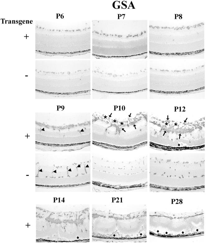
Figure 6.

Histochemical staining for G. simplicifolia lectin (GSA) in transgene-positive (+) and -negative (−) rho/PDGFB1 mice. There is no definite difference in GSA-staining at P6 and P7, but by P8, transgene-positive mice have more stained cells on the surface of the retina. At P9, transgene-positive mice show a multilayered sheet of GSA-positive cells on the surface of the retina. They also show a few positive cells in the inner nuclear layer that could represent penetrating vessels (arrows), but they are much less extensive than the penetrating vessels (arrows) forming the deep capillary beds in transgene-negative mice. At P10 and P12, the superficial, intermediate, and deep capillary beds are seen in transgene-negative mice, but not in transgene-positive mice, which lack identifiable vessels, and instead show a disorganized mass of GSA-positive (arrows) and -negative cells (presumably astrocytes; asterisks) on the surface of the retina and cords of cells invading the retina. Focal areas of traction detachment (diamonds) are seen at P12 and P14, and more extensive detachments are seen at P21 and P28 in transgene-positive mice.
