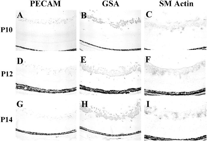
Figure 9.
The increase in vascular cells in rho/PDGFB mice is due to a large increase in pericytes and a much smaller increase in vascular endothelial cells. Rho/PDGFB mice were sacrificed at P10, P12, or P14. Serial frozen sections were stained for platelet-endothelial cell adhesion molecule (PECAM), which selectively stains endothelial cells, G. simplicifolia lectin (GSA), which stains endothelial cells and pericytes, or smooth muscle actin (SM actin), which selectively stains pericytes. At each time point, SM actin staining corresponds closely to GSA staining, with PECAM staining being much less. This suggests that most of the proliferated vascular cells are pericytes.

