
Figure 2.
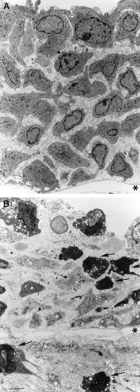
Transmission electron micrographs of the rat common carotid artery 7 days after the induction of IH. Control animal (A): cells with abundant rough endoplasmic reticulum, and oval nuclei. Four hours after PDT (B): ultrastructural features of apoptosis, including nuclear and cytoplasmic condensation, membrane blebbing, cell fragmentation, and apoptotic bodies are noted (arrows). *, internal elastic lamina. Scale bar, 6 μm.