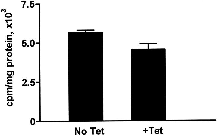
Figure 3.
Analysis of proliferation in MDCK tTA/hPAX2 cells. Cells were grown in the absence or presence of 1 μg/ml of tetracycline and assayed for [3H]-thymidine incorporation as described in Materals and Methods. There is no significant difference between the rate of proliferation in cells with high (No Tet) or undetectable level (+Tet) of PAX2 protein expression. (P ≥ 0.5).

