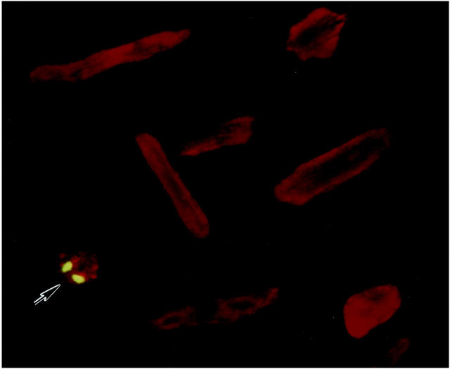
Figure 1.
EMB labeling of a stretched (S) Adp53m-infected myocyte. Green fluorescence reflects EMB-positive nuclei; myocyte cytoplasm shows red fluorescence because of α-sarcomeric actin staining. Several EMB-negative myocytes are illustrated. Confocal microscopy: ×300. n = 4 in all cases. Noninfected myocytes: NS = 0.59 ± 0.10%; S = 0.48 ± 0.16%. AdLacZ-infected myocytes: NS = 0.51 ± 0.19%; S = 0.60 ± 0.15%. Adp53m-infected myocytes: NS = 0.45 ± 0.12%; S = 0.56 ± 0.20%.

