
Figure 3.
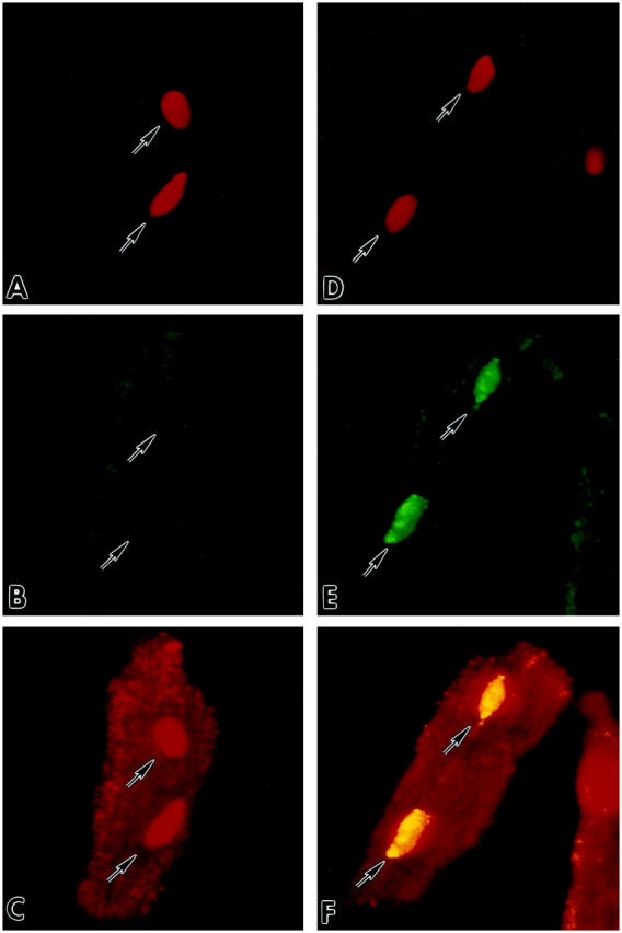
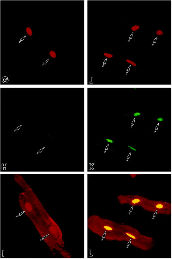
Detection by confocal microscopy of p53 in cultures of AdLacZ- (A–F) and Adp53m- (G–L) infected myocytes before (A–C; G–I) and after (D–F; J–L) stretch. Red fluorescence reflects PI staining of nuclei (A, D, G, J); green fluorescence in B and E corresponds to endogenous wild-type p53 which was detected with anti-rat p53 pAb 246 antibody; p53 labeling was present in the cytoplasm of B and E, and in the nuclei of E only. Green fluorescence in H and K corresponds to Adp53m which was detected with anti-human p53 DO-1 antibody; p53 labeling was present in the cytoplasm of H and K, and in the nuclei of K only. Laser power was set to 90% for images in B and E, and 10% for images in H and K. Red fluorescence depicts α-sarcomeric actin staining of the myocyte cytoplasm in C and F of AdLacZ-infected myocytes, and in I and L of Adp53m-infected myocytes. Red fluorescence of nuclei in C and I illustrates PI labeling alone, and yellow fluorescence of nuclei in F and L illustrates the combination of PI and p53 stainings. Arrows point to unlabeled and labeled nuclei. Confocal microscopy: A–F, ×800; G–L, ×600.